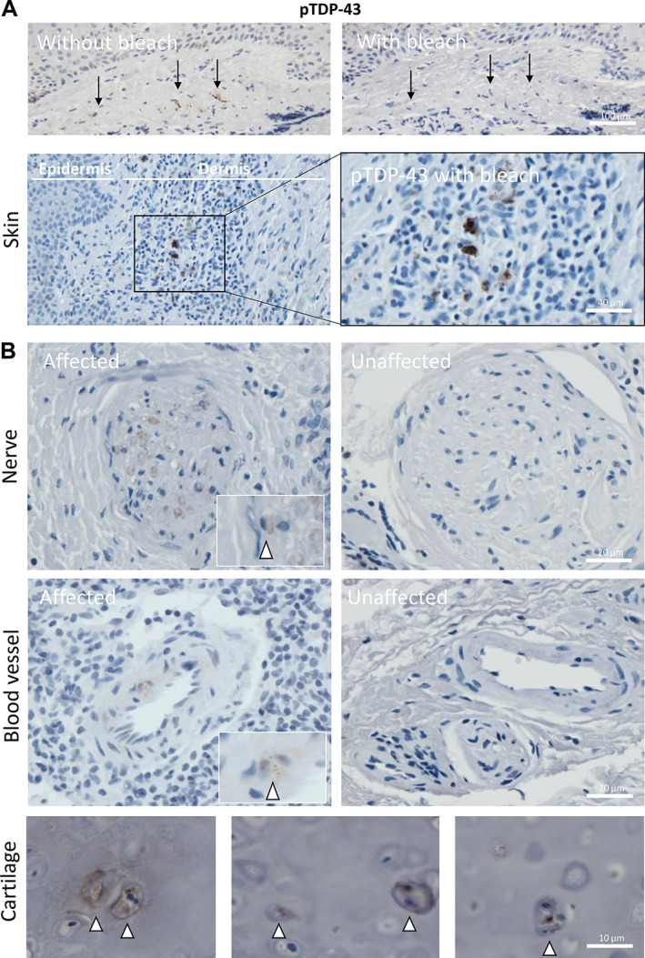
Figure 5.

pTDP‐43 aggregates identified in nerve bundles, endothelial cells, and superficial dermis of skin. (A) Images demonstrating pTDP‐43 staining of skin following removal of brown pigmentation (from melanocytes and/or pigment drop out as a reactive feature) from the dermis and epidermis (above) and pTDP‐43 positive aggregates within the superficial dermis (below). All are taken from patient 26. (B) Example of pTDP‐43 present within peripheral nerves (top left; patient 14) and an example of an unaffected nerve bundle (top right; patient 29) and within endothelial cells of the dermal blood vessels (bottom left; patient 14) and an unaffected blood vessel for comparison (bottom right; patient 29). (C) Three example images taken from patient 26 demonstrating pTDP‐43 aggregates within the chondrocytes of the ear cartilage sampled as part of an excision specimen for a basal cell carcinoma.
