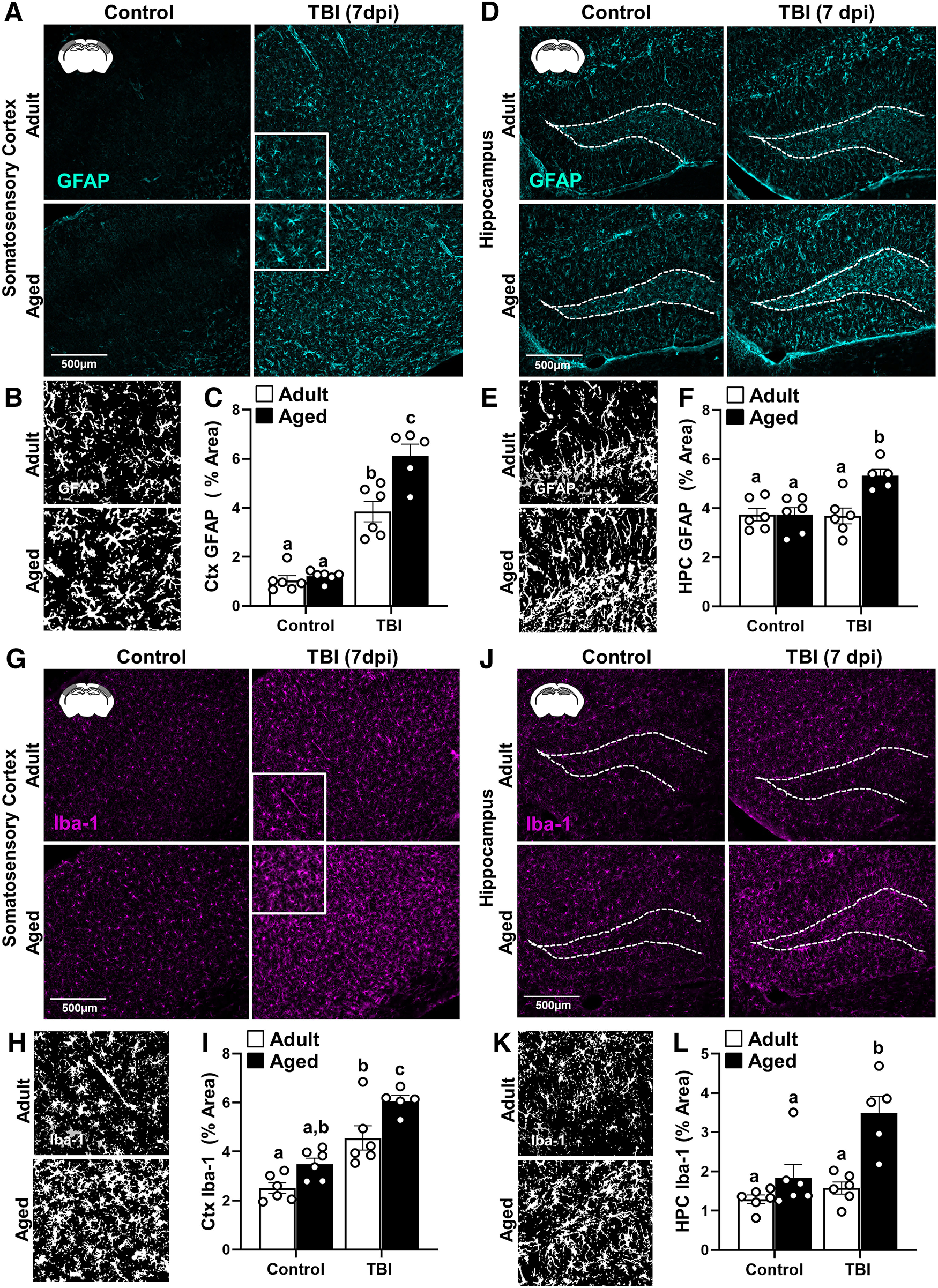
Figure 2.

Amplified morphologic restructuring of astrocytes and microglia in aged mice after TBI. Adult (2 months of age) and aged (18 months of age) male C57BL/6 mice were subjected to midline fluid percussion injury (TBI) or left as uninjured CONs. GFAP+ and Iba-1+ labeling was determined in the SS-Ctx and HPC of adult and aged mice 7 dpi. A, Representative images of GFAP+ labeling in the SS-Ctx. B, Thresholded black and white images of GFAP+ labeling in the SS-Ctx of adult and aged mice 7 dpi. C, Percentage of the area of GFAP+ labeling in the SS-Ctx. D, Representative images of GFAP+ labeling in the HPC. E, Thresholded black and white images of GFAP+ labeling in the HPC of adult and aged mice 7 dpi. F, Percentage of the area of GFAP+ labeling in the HPC. G, Representative images of Iba-1+ labeling in the SS-Ctx. H, Thresholded black and white images of Iba-1+ labeling in the SS-Ctx of adult and aged mice 7 dpi. I, Percentage of the area of Iba-1+ labeling in the SS-Ctx. J, Representative images of Iba-1+ labeling in the HPC. K, Thresholded black and white images of Iba-1+ labeling in the HPC of adult and aged mice 7 dpi. L, Percentage of the area of Iba-1+ labeling in the HPC. Bars represent the mean ± SEM, and individual data points are provided. Means with different letters (e.g., a, b, c) indicate significant post hoc differences between groups (p < 0.05). Groups with the same letter or letters are not significantly different.
