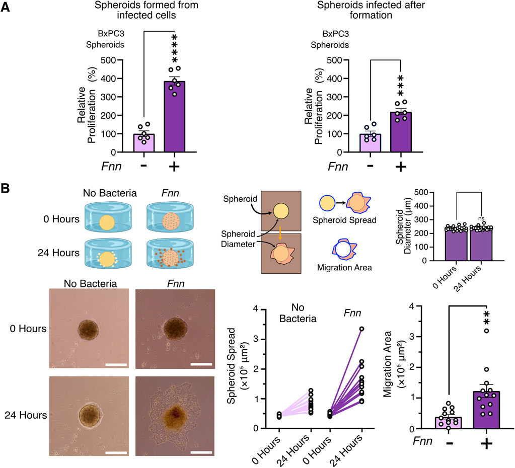
Figure 6: F. nucleatum infection induces proliferation of BxPC3 spheroids and migration of BxPC3 cells from spheroids.
(A) XTT proliferation assays measure proliferation of BxPC3 spheroids (5000 cells) when infected with F. nucleatum at 50:1 MOI both before spheroid formation and after spheroid formation. Data are representative of means ± SEM from N = 6 independent experiments per condition. (B) Representative images of the migration of BxPC3 cells from 5000 cell spheroids in the custom designed chamber. Spheroid diameters were quantified at the start and at the end of the experiment to confirm no expansion and contraction of the spheroids during the duration of the experiment. Migration over 24 hours is quantified by measuring extent of spheroid spread and overall area of migration after normalization to spheroid sizes. Data are representative of means ± SE from N=4 with 3 biological replicates per condition; comparisons by unpaired t-test: * P≤0.05, ** P≤0.01, *** P≤0.001, and **** P≤0.0001; ns, not significant.

