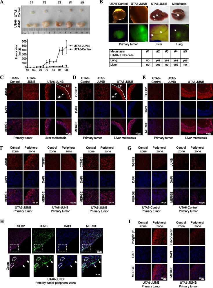
Fig. 7.
Overexpression of JUNB promotes tumor development and metastasis in vivo. A JunB induces tumor growth in immunodeficient mice. UTA6-JUNB and UTA6-control cells were subcutaneously injected in NSG mice. Tumor size in the subcutaneous xenograft model was measured twice a week (lower panel) and 3 months after implantation, mice were euthanized and dissected. Photographs of tumors retrieved at the end of the experiment (upper panel). B JunB induces tumor metastasis in immunodeficient mice. UTA6-control- and UTA6-JUNB-induced primary tumors and GFP expression in these tumors (Left panels, the scale bar is 1 mm), and representative image of liver and lung UTA6-JUNB-induced metastasis and GFP expression in these metastases (right panels, the scale bar is 1 cm). Arrows indicate metastatic lesions. Table reporting EGFP-expressing metastases in lung and liver of the 5 mice xenografted with UTA6-JUNB cells. C–E Higher expression of JUNB, CCNE1, and TGFB2 in UTA6-JUNB liver metastases than in primary UTA6-JUNB tumors. Representative immunofluorescence images of paraffin-embedded UTA6-JUNB and UTA6-Control primary tumor tissues, and UTA6-JUNB liver metastases. C Corresponds to JUNB, D corresponds to CCNE1, and E corresponds to TGFB2 analyses, respectively. DAPI staining (blue) indicates cell nuclei. Scale bars are indicated. F JUNB and TGFB2 levels are increased in the border of UTA6-JUNB expressing primary tumors. Representative immunofluorescence images of the central and peripheral zones of UTA6-JUNB. JUNB, TGFB, CCNE1 (red), and cell nuclei (blue) are observed. G JUNB and TGFB2 levels are similar in the central and peripheral zones in control tumor tissues. Representative immunofluorescence images of the central and peripheral zones of UTA6-Control tumor tissues. JUNB, TGFB (red), and cell nuclei (blue) are observed. H JUNB and TGFB colocalization in the UTA6-JUNB primary tumor border. Representative immunofluorescence images of UTA6-JUNB primary tumor tissue. TGFB2 (magenta), JUNB (green), and cell nuclei (blue) are observed. Arrows indicate cells co-expressing JUNB and TGFB2 proteins. I Integrin β1 and Fibronectin levels are increased in the border of UTA6-JUNB expressing primary tumors. Representative immunofluorescence images of UTA6-JUNB and control primary tumor tissues. Integrin β1 (red, left panel), fibronectin (red, right panel), and cell nuclei (blue) are shown. Scale bars are indicated. LT: Liver Tissue MT: Metastatic tissue

