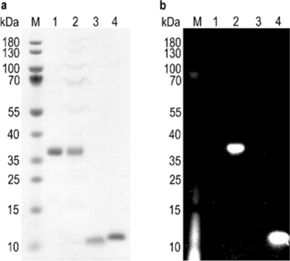
Fig. 2.

Confirmation of AzK incorporation in Stx1B K9AzK and scFv OKT3 E129AzK by fluorophore labelling. The purified proteins scFv OKT3 (lane 1, MWcalc 30 kDa), scFv OKT3 E129AzK (lane 2, MWcalc 30 kDa), Stx1B (lane 3, MWcalc 9 kDa) and Stx1B K9AzK (lane 4, MWcalc 9 kDa) were incubated with DBCO-Cy3 as detailed in the materials and methods section and then analyzed on a 4–12% SDS gel. a`Proteins stained with Coomassie protein stain. b SDS gel irradiated at 635 nm before staining with Coomassie protein stain. The sizes of the molecular weight marker (M) bands are indicated on the left margin of the gels
