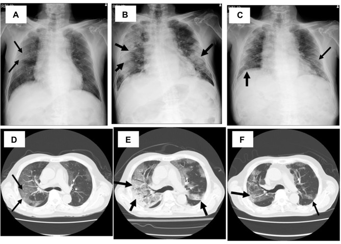
Figure 2.
Chest X-ray (A–C) and computed tomography (D–F) of the case 2 patient on day 10 (A and D), day 15 (B and E), and day 30 (C and F). GGOs are found in both lung fields on day 10 (A and D). They were worse and lesions, including the densities were increased on day 15 (B and E), but they have finally improved on day 30 (D and F). Arrows indicated the abnormal shadows in chest X-ray and CT, such as interstitial shadows and GGO.

