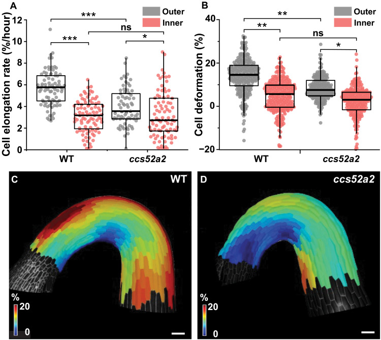
Fig. 2. Endoreplication mediates in control of differential cell elongation and mechanical properties during hook development.
(A) Cell elongation rate of epidermal cells (%/hour) at 0 to 400 μm from the shoot apex in the WT (n = 5; outer, 80 cells; inner, 88 cells) and ccs52a2 (n = 6; outer, 69 cells; inner, 84 cells). (B) Quantification of the deformation of the epidermal cells in the WT (n = 6; outer, 240 cells; inner, 250 cells) and ccs52a2 (n = 6; outer, 197 cells; inner, 213 cells). Thick lines in the boxes indicate medians. (C and D) Heatmaps of cell deformation following osmotic treatment for the WT (C) and ccs52a2 (D). Statistical significance was calculated using Student’s t test (unpaired, two-tailed) and is indicated as follows: ns, *P < 0.05, **P < 0.01, and ***P < 0.001. Scale bars, 100 μm.

