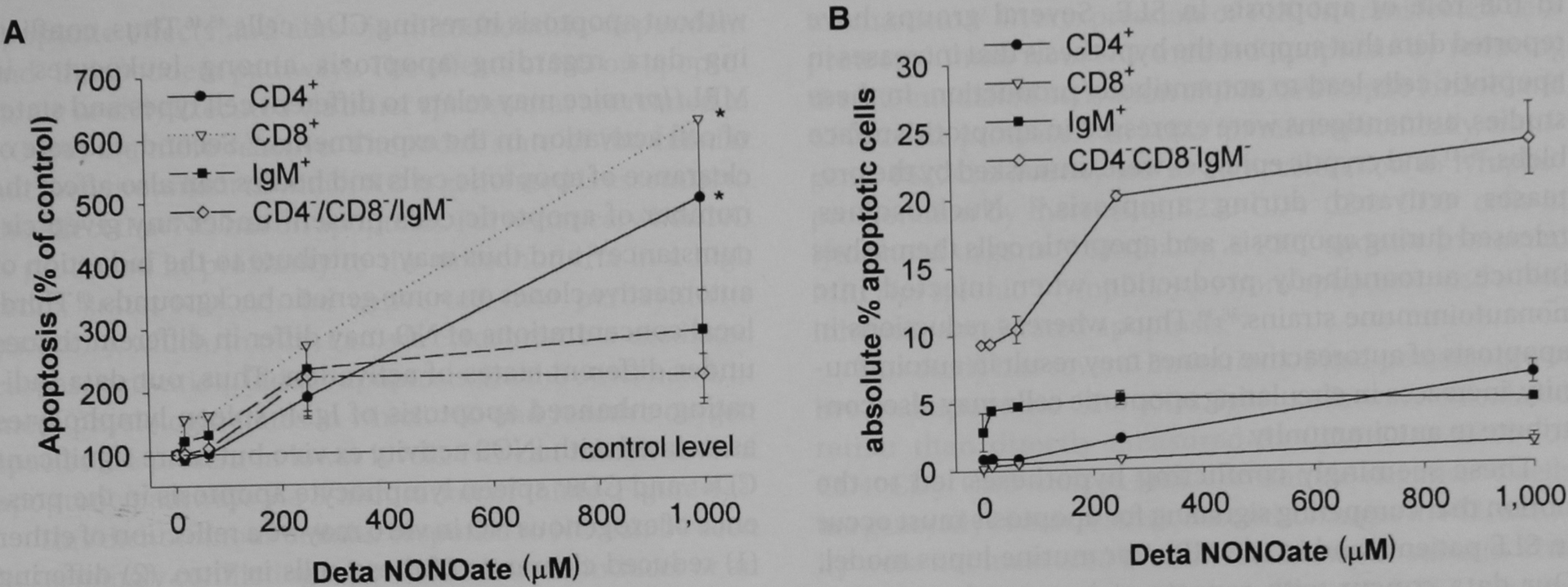
FIGURE 3.

Apoptosis of MRL/lpr spleen lymphocyte subsets with exogenous nitric oxide in vitro. MRL/lpr spleen lymphocytes were isolated and cultured overnight in varying concentrations of Deta NONOate. Cells were harvested and analyzed for apoptosis using flow cytometry with annexin V–fluorescein isothiocyanate and phycoerythrin cell surface marker (CD4+, CD8+, and IgM+) staining. A, The results were normalized to the percentage of apoptotic cells relative to control wells (not exposed to Deta NONOate) ± standard error and were calculated from the average of five experiments. Apoptosis of subsets was reported as follows: CD4+ cells as closed circles, CD8+ cells as open inverted triangles, IgM+ cells as closed squares, and CD4−CD8−IgM− cells as open diamonds. Deta NONOate induced significant apoptosis among CD4+, CD8+, and CD4−CD8−IgM− cells (asterisk signifies p < .05). B, The results of a representative single experiment reported as the percentage of apoptotic intact cells in each subset. Subset analysis was not performed for BALB/cJ spleen lymphocytes.
