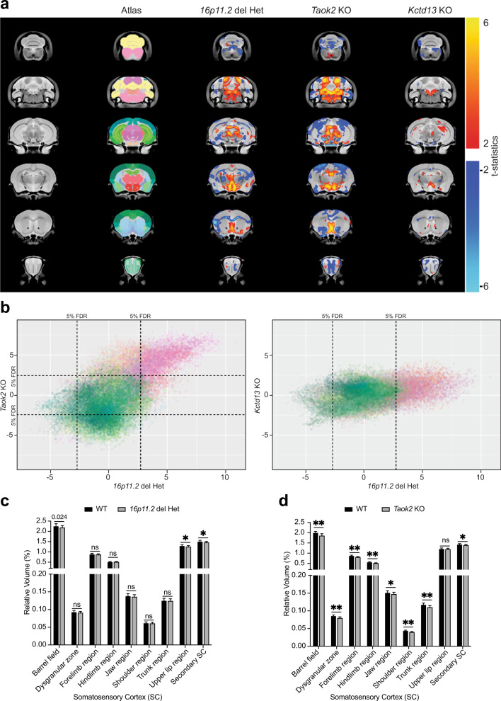
Fig. 7. Alterations in brain anatomy in the 16p11.2 microdeletion mouse model resemble the phenotype of Taok2 KO mice but not of Kctd13 KO mice.
a Het 16p11.2 microdeletion mice and Taok2 KO mice have altered brain morphology. A voxelwise analysis highlighting significant differences in relative volume (images show the lowest threshold of 5% false discovery rate (FDR) for Het 16p11.2 deletion mice, Taok2 KO mice and Kctd13 KO mice) throughout the brain between the respective WT and deletion mice. T-stats indicate positive or negative changes compared with the respective WT littermate brains. Taok2 KO mice but not 16p11.2 deletion or Kctd13 KO mice had increased absolute brain volume compared with WT mice (Taok2 WT = 16, KO = 23; 16p11.2 WT = 14, Het = 15; Kctd13 WT = 23, KO = 23 mice from three different cohorts, statistics by linear model. b Correlation analysis of relative volumes of brain regions in Het 16p11.2 deletion mice and in Taok2 KO mice revealed strong positive associations between Het 16p11.2 deletion and Taok2 KO mice but not between Het 16p11.2 deletion and Kctd13 KO mice; each point in the plot indicates a voxel in the brain, and the color indicates the anatomical area from which the voxel originated (colors from the Allen atlas, indicated in Column 2 of a). c, d The somatosensory cortex is particularly affected in Het 16p11.2 deletion mice and in Taok2 KO mice (statistics by linear model corrected for multiple comparisons using FDR; values are the mean ± S.D.; see Supplementary Table 1 for FDR and p values).

