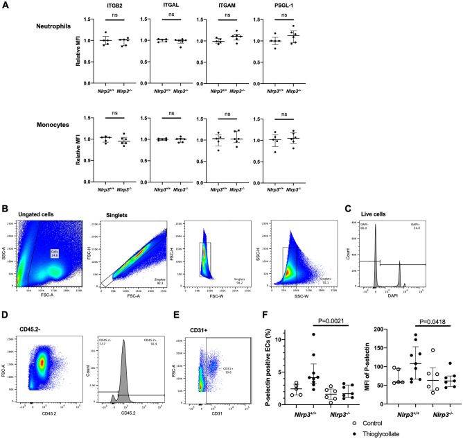
Figure 4.
Nlrp3 deficiency reduces membrane expression of P-selectin on the inflamed endothelial cells, while circulating leukocyte adhesion receptors expression is not altered. Neutrophils and monocytes in peripheral blood were collected from Nlrp3+/+ and Nlrp3−/− mice 4 h after injection of thioglycollate broth (n = 5:6). (A) Surface expressions of integrins; integrin β2 (ITGB2, CD18), integrin αL (ITGAL, CD11a), integrin αM (ITGAM, CD11b), and P‐selectin glycoprotein ligand 1 (PSGL-1, CD162) were analyzed by flow cytometry. The relative MFI was calculated as a relative value to an average MFI of Nlrp3+/+. (B-F) Endothelial cells were isolated from the mesentery of Nlrp3+/+ and Nlrp3−/− mice 4 h after injection with thioglycollate (Thioglycollate, closed circles, n = 9:7) or PBS (Control, open circles, n = 5:6). Surface P-selectin expression was analyzed by flow cytometry. (B–E) Gating strategy for isolated endothelial cells. Endothelial cells were defined as negative for CD45.2 and positive for CD31; (B) ungated cells and singlets, (C) live cells as negative for DAPI, (D) CD45.2 negative and (E) CD31 positive. (F) P-selectin expression on gated endothelial cells. Left: percentage of P-selectin positive endothelial cells. Right: mean fluorescent intensity (MFI) of P-selectin.

