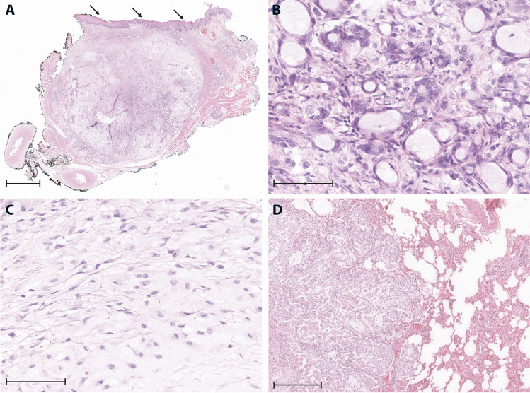
Fig. 2.
A Overview (1.4 × ; scale bar: 500 µm) of the resection specimen, showing a largely well-circumscribed, focally infiltrative, and variably cellular, submucosal tumor (arrows: mucosa). B High-power magnification (40 ×) from cell-rich area with monomorphic, unilayered tubules and microcysts with bland cytological features. C Cell-poor area with monomorphic spindle cells, representing fibroblastic cells or modified myoepithelial cells (40 × ; scale bar: 40 µm). D Metastatic nodule in the lung with cell-rich tumor component (10 × ; scale bar: 100 µm)

