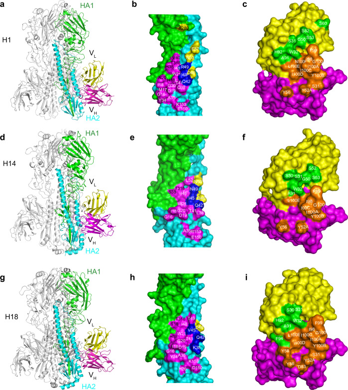
Fig. 3. Structures of the PN-SIA28/H1, PN-SIA28/H14, and PN-SIA28/H18 complexes.
The overall structures of the PN-SIA28/H1 (a), PN-SIA28/H14 (d), and PN-SIA28/H18 (g) complexes are displayed in cartoon representation. The PN-SIA28 Ab binds the conserved stem regions of the H1, H14, and H18 HAs. The epitope residues of PN-SIA28 in HA1 and HA2 of H1 (b), H14 (e), and H18 (h) are denoted in black and white characters, respectively. Residues of HA that are in contact with the heavy chain of PN-SIA28 are colored magenta, residues that are in contact with the light chain of PN-SIA28 are colored yellow, and residues that are in contact with both chains of PN-SIA28 are colored blue (b, e, h). The residues of PN-SIA28 responsible for the HA binding in the PN-SIA28/HA complexes (c, f, i) are marked in white characters. The heavy chain is colored magenta, and the light chain is colored yellow. The residues contacting the HA are colored orange for the heavy chain and colored pink for the light chain (c, f, i).

