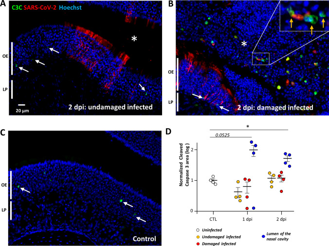
Fig. 1.
Apoptosis occurs in desquamated cells in the lumen of the nasal cavity following SARS-CoV-2 infection but not in the olfactory epithelium. Representative images of an infected intact (A), infected damaged (B) area of the olfactory epithelium at 2 days post-infection (dpi) and in a control animal (C). Apoptotic cells in the olfactory epithelium are indicated by a white arrow (OE; olfactory epithelium/LP lamina propria). The lumen of the nasal cavity is indicated by a white asterisk and is filled with cells, some of which colocalize in their nucleus cleaved caspase 3 signal (orange arrow). (D) Cleaved caspase 3 signal in the olfactory epithelium normalized to control (log 10, Mean ± SEM, n = 4, *p < 0.01 (Mann–Whitney test))

