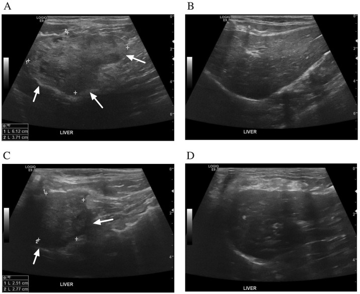
Figure 3.
Ultrasonographic findings of the liver after treatment with prednisolone. (A,B) 12 weeks after prednisolone treatment. (A) The left lobe of liver showed a poorly defined mass (arrows) containing small irregular anechoic cavitations. (B) The right lobe of liver had a homogeneous parenchyma. There was no evidence of intrahepatic bile duct dilatation. (C,D) Twenty-three months after stopping prednisolone. (C) The left lobe of liver showed a heterogenous mass (arrows). (D) The right lobe of liver had a mild heterogenous parenchyma and round border. There was no evidence of common bile duct and intrahepatic bile duct dilatation.

