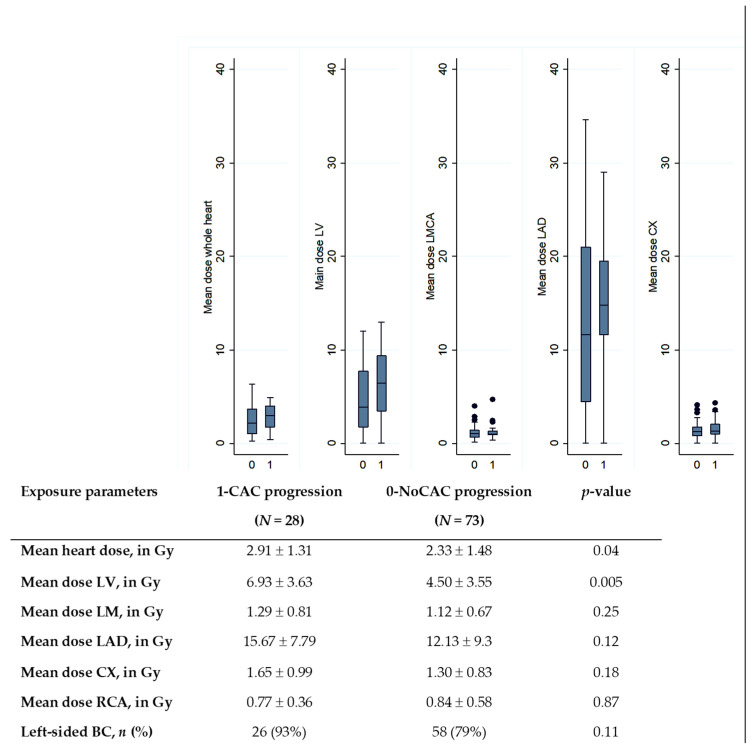
Figure 1.
Comparison of cardiac doses according to the CAC progression status (0 for no CAC progression; 1 for CAC progression). LMCA: left main common coronary artery, LAD: left anterior descending artery, CX: circumflex artery; RCA: right coronary artery. The central value of the box indicates the median, the borders of the box indicate the quartiles (25th and 75th), and the extremities indicate the minimum and maximum values. p-value: results of Wilcoxon test to compare dose distributions.

