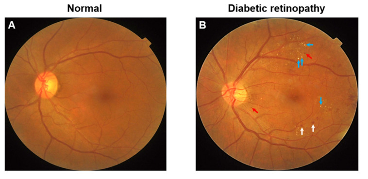
Figure 1.
(A) Color fundus photograph of a diabetic individual without retinopathy. (B) Color fundus photograph of a diabetic individual with signs of moderate non-proliferative diabetic retinopathy. Notably, features include microaneurysms (red arrows), dot-and-blot hemorrhages (white arrows), and hard exudates (blue arrows, HE).

