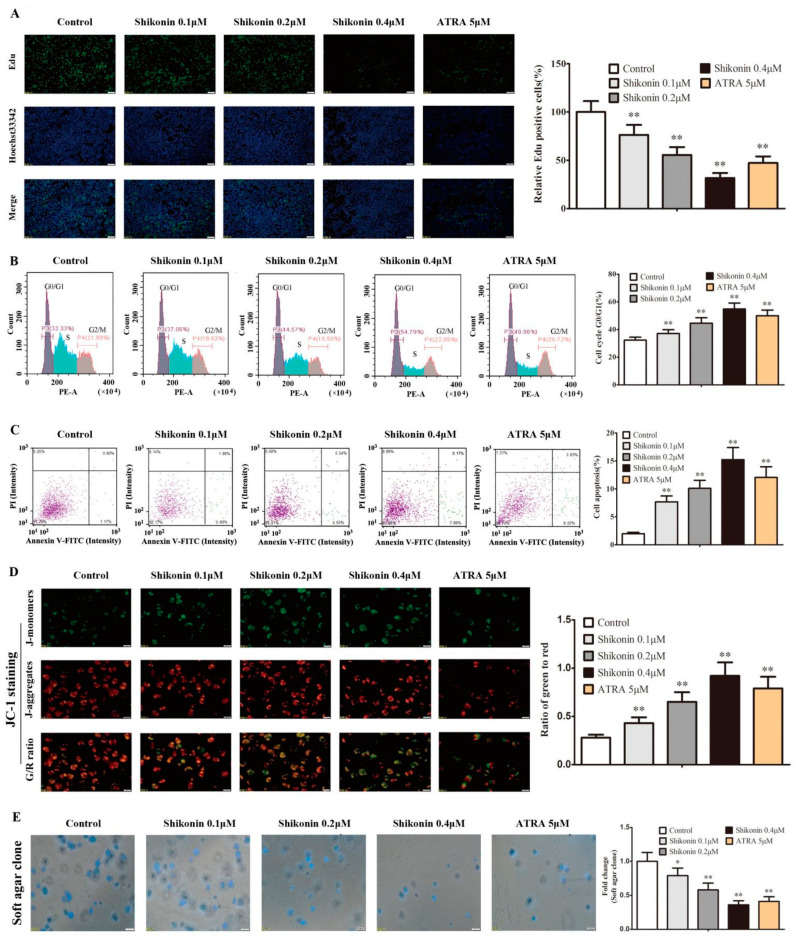
Figure 6.
Shikonin-differentiated HL-60 cells gradually lose the ability of cell proliferation and self-renewal. HL-60 cells were treated with shikonin for 10 d. (A) Cell proliferation was detected by EdU incorporation. (B) The cell cycle was detected by PI staining using flow cytometry. (C) Cell apoptosis was detected by Annexin V-FITC/PI staining using flow cytometry. (D) The mitochondrial membrane potential (MMP) was detected by JC-1 staining, which forms J-monomers with green fluorescence to label the low MMP and J-aggregates with red fluorescence to label the high MMP. (E) Cell self-renewal was detected by soft agar clone formation. The data were presented as the mean ± SD (n = 3). * p < 0.05; ** p < 0.01 compared to the untreated control.

