Figure 1.
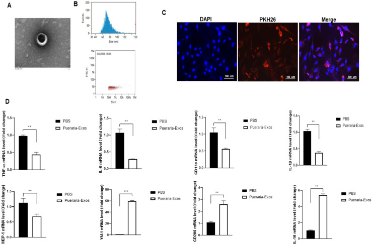
Characterization of exosomes isolated from P. lobata. (A) Transmission electron microscopy (TEM) images of exosomes from P. lobata. Scale bar, 100 nm. (B) Particle size distribution of P. lobata-derived Exos measured by dynamic light scattering. (C) Fluorescence microscopy images of macrophages co-cultured with PKH67-labeled P. lobata-derived Exos for 6 h. All scale bars = 100 μm. (D) The gene profiles of the M1 (IL-6, IL-1β, TNF-α, MCP-1, and CD11c) and M2 (IL-10, YM1, and CD206) phenotypes as assessed by qPCR from macrophages in vitro. Data were normalized by the amount of 18s mRNA and expressed relative to the corresponding PBS. Experiment was performed in duplicates 3 independent times. ** p < 0.01; *** p < 0.001. Data are shown as means ± SEM.

