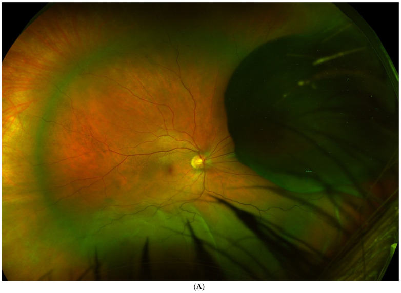
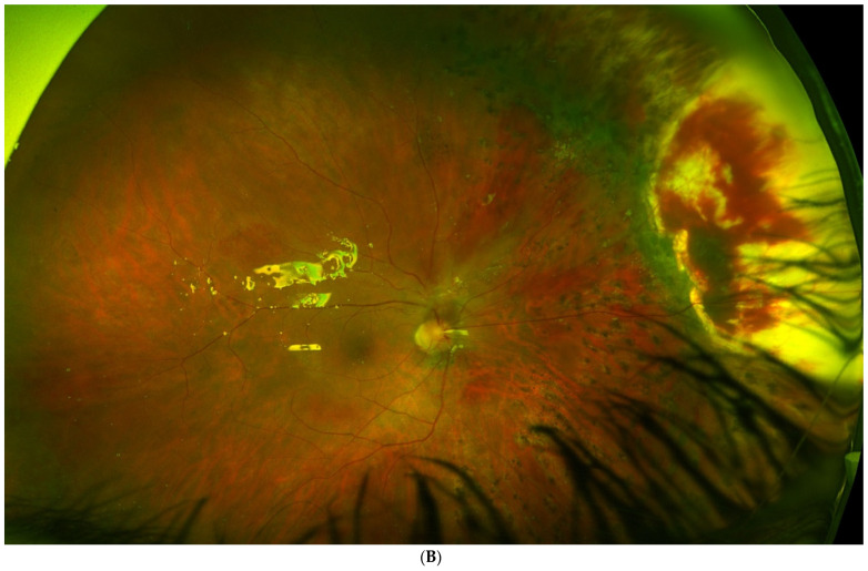
Figure 2.
(A). Tumour located in the preequatorial and superior nasal quadrant, with extensive inferior retinal detachment, in which a partial lamellar scleral resection was performed. (B). Fundus image shows the extensive surgical coloboma, as well as the perilesional photocoagulation and retinal reattachment.


