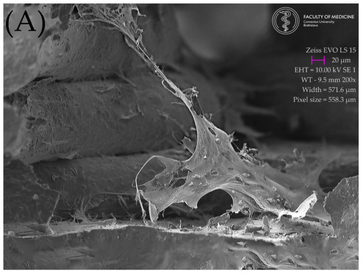
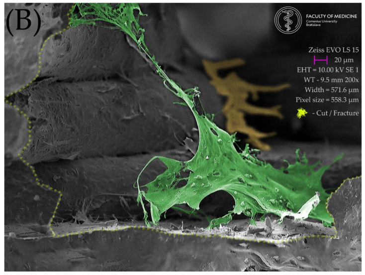
Figure 9.
Lateral view of the interior of the scaffold floor, created with an ultra-sharp razor blade. A large flat cell in the foreground shows exceptionally long cell projections firmly attached to the lateral surfaces of the scaffold. The openings between the scaffold lines appear to be a friendly environment for cell growth: (A) original SEM image; (B) enhanced image showing the scaffold section and the green-colored cell attached to the scaffold from bottom to the top, with a yellow-colored cell in the background climbing up in the posterior corner of the scaffold.


