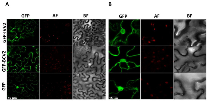
Figure 5.
Subcellular localization of V2 from beet curly top Iran virus fused to GFP in epidermal cells of wild type Nicotiana benthamiana. (A) Leaves were agroinfiltrated with a construct expressing the GFP (GFP), GFP-V2 fusion protein from beet curly top Iran virus (GFP-IVV2), or the GFP-V2 fusion protein from beet curly top virus (GFP-BCV2). Samples were observed under the confocal microscope at 36 h post infection. (B) Close up confocal images of the observed areas. GFP fluorescence (GFP), autofluorescence (AF), and the bright field channel (BF) are shown.

