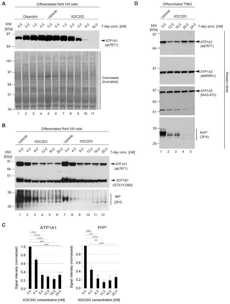
Figure 6.
KDC203 reduces steady-state PrPC levels in human neural cell lines. (A) Oleandrin and KDC203 reduce steady-state ATP1A1 protein levels to a similar degree. Side-by-side Western blot-based comparison of ATP1A1 signal intensities in cellular extracts derived from ReN VM cells following 7-day treatment with the respective CGs (equal amount of total protein loaded). Because the Western blot was not stripped of the ATP1A1-directed primary and secondary antibodies, before it was stained with Coomassie, a stronger signal can be seen at the 85–90 kDa level, where the ATP1A1 detection antibodies bound. (B) 7-day exposure of ReN VM cells does not only affect ATP1A1 protein levels by also causes a concentration-dependent reduction in the steady-state protein levels of the NKA β subunit ATP1B1 and PrPC. Note that the total amount of protein loaded in the two biological replicate series was not identical for the PrP-directed 3F4 blot; to capture an informative linear range, half the amount of total protein was loaded for samples shown on the right hand side. (C) Quantitation of Western blot signal intensities of ATP1A1 and PrPC following 7-day KDC203 treatment of ReN VM cells at concentrations indicated, with each value being computed from the analysis of three biological replicates. (D) The KDC203-dependent reduction in steady-state PrPC protein levels is not an idiosyncrasy of ReN VM cells but can also be observed in other neural cell models, including differentiated human glioblastoma cells (T98G). Asterisks indicate the level of statistical significance, i.e., one asterisk denotes p < 0.05, with each additional asterisk denoting tenfold lower p-values.

