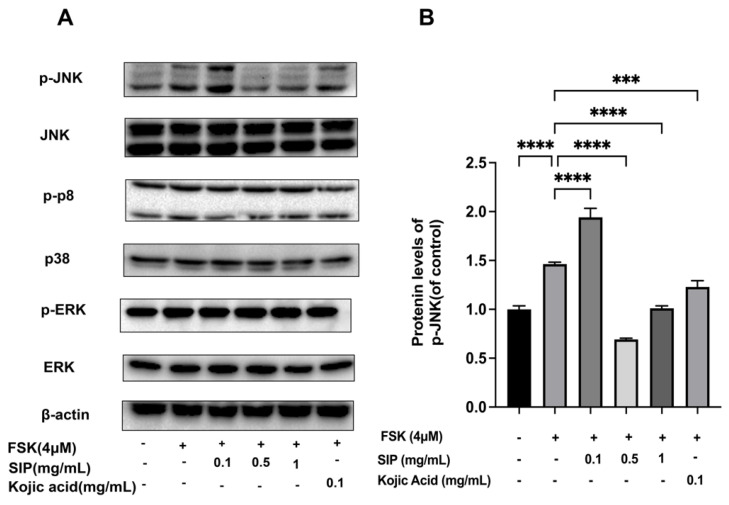
Figure 7.
Effects of SIP on MAPK signaling pathway in FSK-stimulated B16F10 melanoma cells. (A) Phosphorylation and total Protein expression levels of ERK, p38, and JNK were detected by Western blot following SIP treatment with the indicated concentration. (B) The densitometric analysis of p-JNK using Photoshop software. Results were presented as mean ± SD. Statistical analysis was performed with one-way ANOVA, followed by Tukey’s multiple comparison test (n = 3; *** p < 0.001, and **** p < 0.0001 compared with the forskolin-stimulated group).

