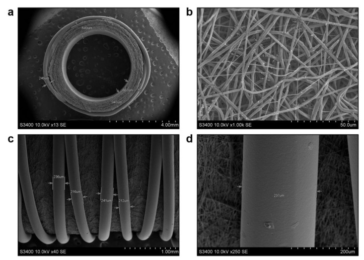
Figure 1.
Electron microscopy examination of electrospun PCLIlo/CAD TEVGs before their implantation. (a) Tubular scaffold with a highly porous wall and anti-aneurysmatic PCL sheath, ×13 magnification, scale bar: 4 mm; (b) Graft wall consisting of micro- to nanoscale fibers and interconnected pores, ×1000 magnification, scale bar: 50 µm; (c) Large, continuous and partially crossed filaments of an anti-aneurysmatic PCL sheath, ×40 magnification, scale bar: 1 mm; (d) A close-up of the filament within an anti-aneurysmatic PCL sheath, ×250 magnification, scale bar: 200 µm. Accelerating voltage: 10 kV.

