Figure 11.
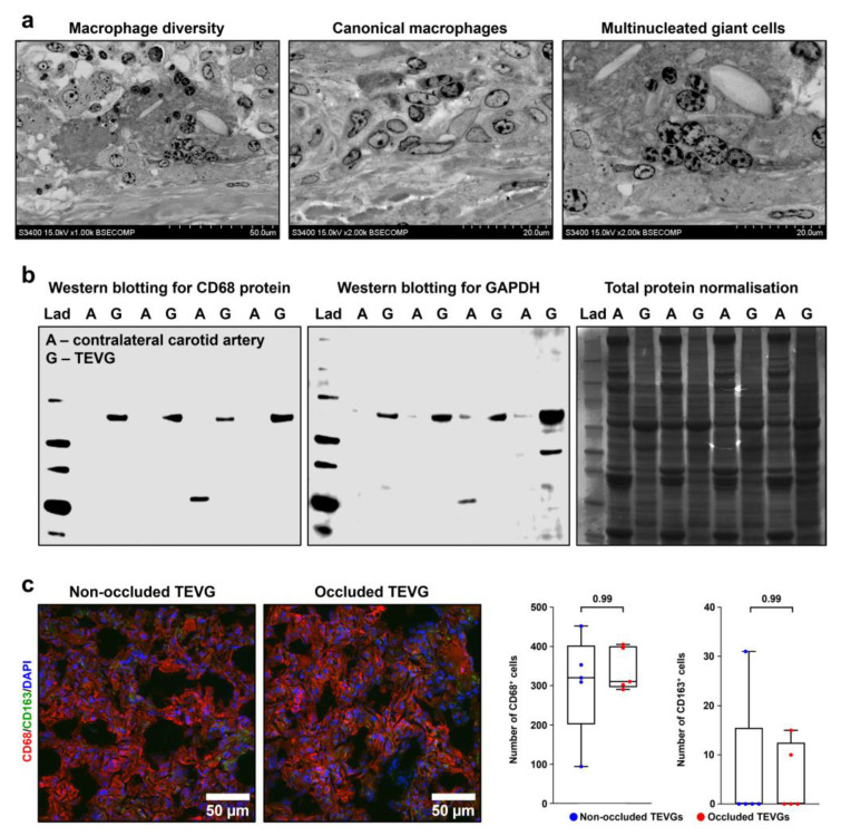
TEVGs but not intact contralateral carotid arteries are infiltrated by macrophages. (a) Backscattered scanning electron microscopy (EM-BSEM) shows massive macrophage invasion within the tunica adventitia of the TEVG. Left: overview of invading macrophages; centre: canonical macrophages; right: multinucleated giant cells. Magnification: ×1000 (left) and ×2000 (centre and right), scale bar: 50 and 20 µm respectively, accelerating voltage: 15 kV. (b) Western blotting of TEVG and contralateral carotid artery lysate for CD68 protein, representative image (left) and for glyceraldehyde 3-phosphate dehydrogenase (GAPDH), representative image (centre), and loading control by Coomassie Brilliant Blue staining (right). (c) Immunofluorescence staining for the pan-macrophage marker CD68 (red colour) and M2 macrophage marker CD163 (anti-inflammatory and pro-fibrotic macrophage specification, green colour). Magnification: ×400, scale bar: 50 µm. Nuclei are counterstained with DAPI. Representative images (left) and semi-quantitative analysis (right). Each dot on the plots represents the measurement from one image (n = 5 images per group). Whiskers indicate the range, box bounds indicate the 25th–75th percentiles, and centre lines indicate the median. p values are provided above boxes, Mann–Whitney U test.

