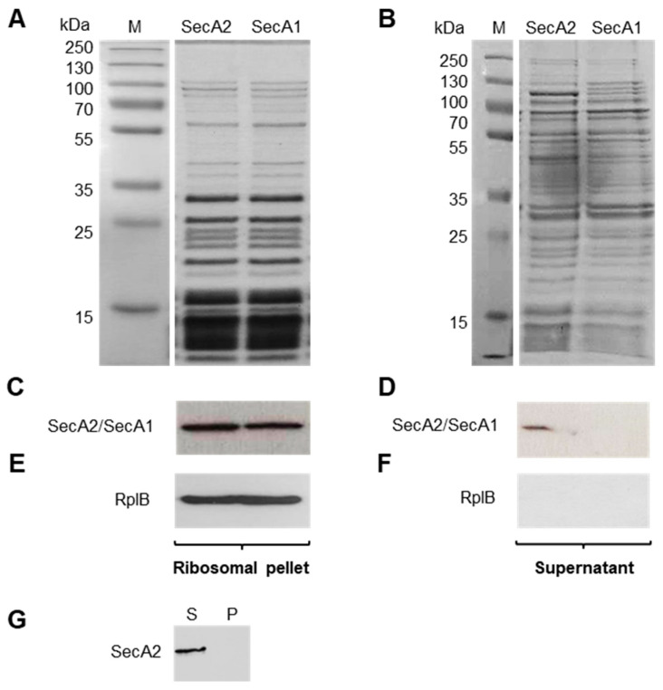
Figure 3.
Co-sedimentation experiments of SecA2 with ribosomes. Purified SecA2 and SecA1 proteins (1 µM) were mixed with purified ribosomes (4 µM) and sedimented by sucrose cushion ultracentrifugation. Coomassie stained gel of ribosomal pellet (A) and the corresponding supernatant (B) are depicted. Western blot analyses were used to detect SecA2 and SecA1 in the ribosomal pellet (C) and in the supernatant (D); (E,F) detection of the ribosomal protein RplB in the ribosomal pellet and the supernatant; and (G) SecA2 sedimentation in the pellet (P) and supernatant (S) fractions in the absence of ribosomes.

