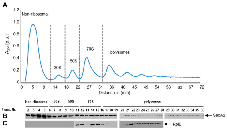
Figure 4.
Detection of SecA2 on polysomes. (A) the ribosome profile of Lm-∆secA2::SecA2strep is depicted. The first gradient fractions show free ribosome-unbound low molecular weight proteins. The ribosomal components 30S, 50S, 70S and polysomes were detected at higher sucrose concentration; and (B,C) Detection of SecA2 and the ribosomal protein RplB in different gradient fractions. Ribosomal peaks were normalized to the same gradient baseline by subtracting the area under (a.u.) the polysome profile for quantitative comparison.

