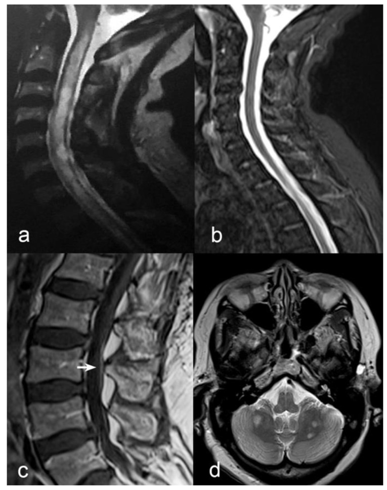
Figure 1.
(a) Sagittal T2-weighted MRI of the cervical spine in the acute phase showing heterogeneous hyperintensity from C2 to the first thoracic segments with predominant central involvement. (b) 1-year after onset, sagittal STIR-T2-weighted MRI of cervical spine showed absence of previously seen inflammatory lesions. (c) Contrast enhancement of cauda equina roots (white arrow) was observed in sagittal T1-weighted sequences after Gadolinium administration. (d) Multiple bilateral round-shaped inflammatory lesions in cerebellar white matter were evident on axial T2-weighted axial scan (d).

