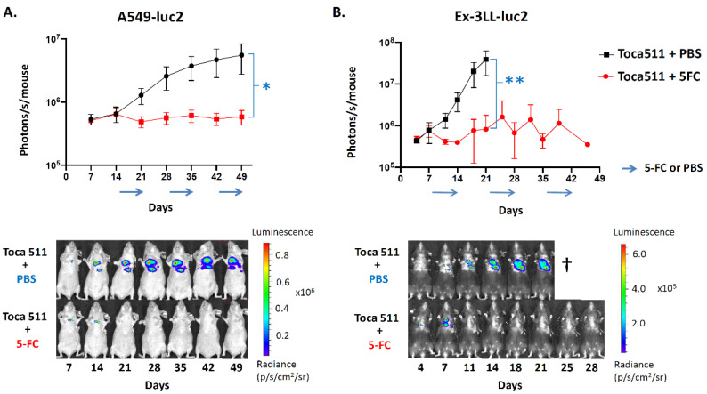
Figure 8.
Bioluminescence imaging of Toca 511/5-FC prodrug activator gene therapy in orthotopic pleural dissemination models of lung cancer. Luciferase-marked human A549-luc2 (A) or murine Ex-3LL-luc2 (B) tumors were established by intrathoracic injection and Toca 511 infection as described in Methods, and IVIS optical imaging was performed at serial time points (n = 5 per group). (A) In the A549-luc2 model, 7-day cycles of 5-FC (500 mg/kg, red line) or PBS (black line) were administered every other week (arrows) from day 14 post-tumor establishment; * p = 0.0147. (B) In the Ex-3LL-luc2 model, 7-day cycles of 5-FC (500 mg/kg, red line) or PBS (black line) were administered every other week starting from day 7; ** p = 0.0326. Lower panels show representative serial imaging results from an individual animal in each treatment group for each model.

