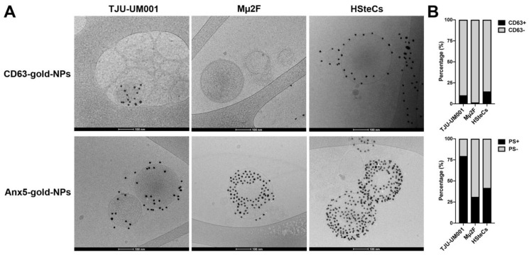
Figure 2.
Morphology of UM-derived EVs. (A) Large EVs isolated from metastatic UM cell lines TJU-UM001 and Mµ2F or HSteCs (healthy liver cell controls) were labeled with 10-nm CD63-gold-NPs or Anx5-gold-NPs (dark particles) for cryo-TEM analysis. Scale bars, 100 nm. (B) The percentage of EVs positive for CD63 or PS was quantified for each cell type.

