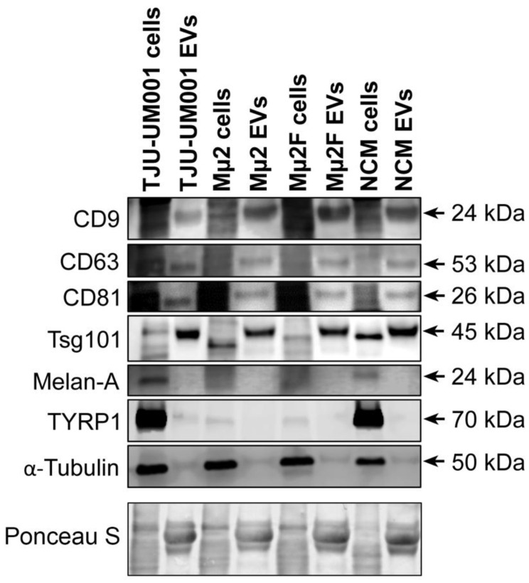
Figure 3.
Exosomal and melanocytic markers present in UM-derived EVs. Cell pellets and EV fractions isolated from UM cell lines TJU-UM001 (metastatic), Mµ2 (non-metastatic), and Mµ2F (metastatic), or NCMs, were analyzed for their expression of exosomal (CD9, CD63, CD81, Tsg101) or melanocytic (Melan-A, TYRP1) markers by Western blotting. The anti-α-tubulin was used as marker of cytosolic proteins. Ponceau S staining was used as a loading control. Arrows indicate the specific band for each marker.

