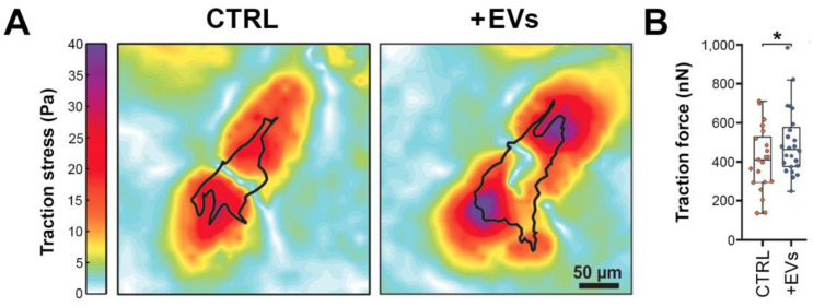
Figure 6.
Increased contractility of stellate cells after the internalization of UM-derived EVs. HSteCs were grown on 5 kPa polyacrylamide gels containing fluorescent beads and exposed for 24 h to large EVs from metastatic UM cells TJU-UM001. Then, bead displacements between stressed (+EVs) and null (CTRL) states were measured using TFM. (A) Representative traction stress maps (Pa) and (B) traction force (nN) are shown for untreated (CTRL) or treated (+EVs) HSteCs. Scale bar, 50 µm. Plots are median ± the minimum and maximum values. * p < 0.05 (Welch’s t-test).

