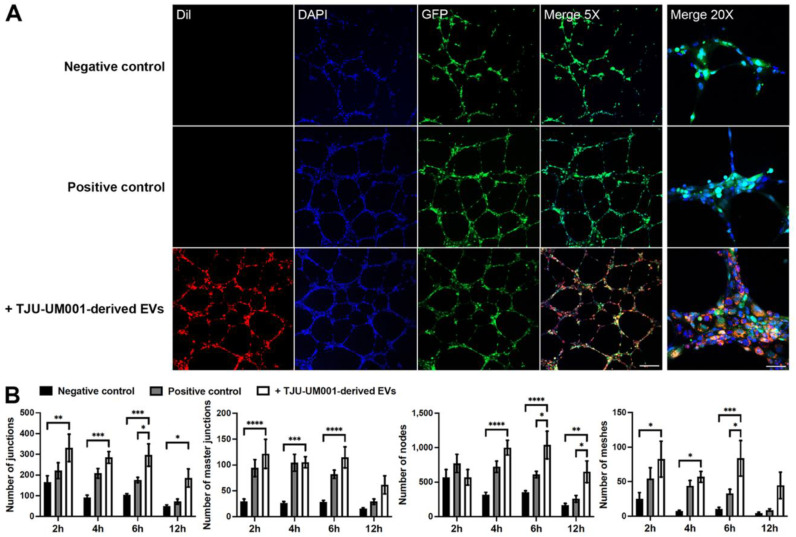
Figure 8.
UM-derived EVs increased the formation of capillary-like tubes by endothelial cells. (A) Untreated GFP-HUVECs (green) were cultured for 12 h on Matrigel® with medium without VEGF (negative control; top panels) or medium supplemented with VEGF (positive control; middle panels). Images are from the 6 h time point. Nuclei were counterstained with DAPI (blue). Scale bars: Merge 5X, 200 µm; Merge 20X, 50 µm. (A) Large EVs isolated from metastatic UM cell line TJU-UM001 were labeled with DiI (red) and added to GFP-HUVECs (green) for 24 h before their seeding on Matrigel® for 6 h (bottom panels). (B) The number of junctions, master junctions, nodes, and meshes were quantified in function of time (hours) for each condition. Error bars indicate SEM (n = 3); * p < 0.05, ** p < 0.01, *** p < 0.001, and **** p < 0.0001 (two-way ANOVA with Sidak’s multiple comparisons test).

