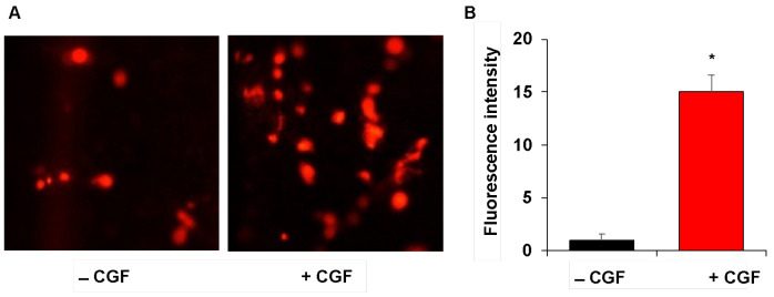
Figure 3.
Endothelial cell adhesion on CGF-permeated implants. Implants without/with CGF (−/+ CGF) were incubated in the presence of DilC18-labeled endothelial cells and their adhesion was monitored by a fluorescence microscope (×100 magnification) (A). The fluorescence intensity was quantified by ImageJ software and reported as arbitrary units (B). The results are expressed as the means ± SD of triplicate measurements from three independent experiments. (* p < 0.05 versus implants without CGF).

