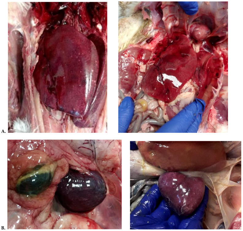
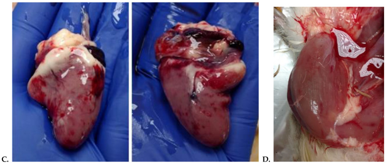
Figure 1.
(A) disseminated, white necrotic spots in an enlarged and congested liver; (B) enlarged and congested spleen with clear foci suggestive of necrosis; (C) epicardial multifocal to coalescing haemorrhage on the surface of the heart muscle; (D) Leg muscle haemorrhages and subcutaneous oedema from a GRV infected goose.


