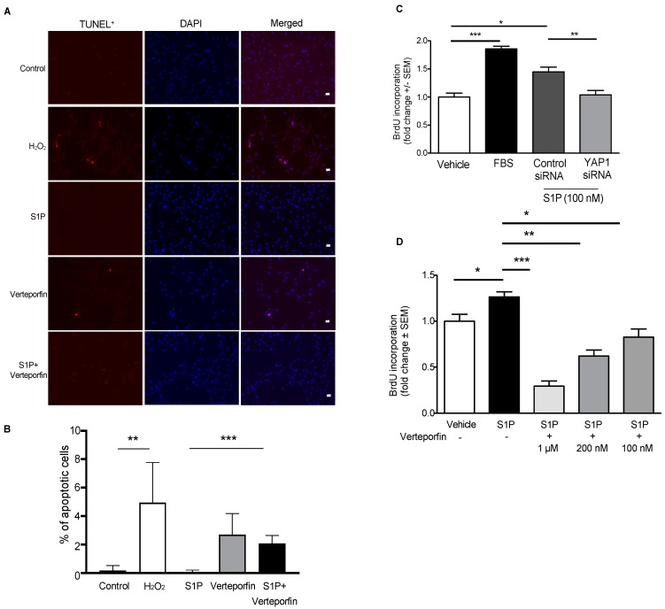
Figure 9.
YAP1 regulates S1P-mediated proliferation in hPASMCs. (A) Representative images and (B) quantitative data from TUNEL assays. hPASMCs were cultured on a coverslip in a six-well cell culture plate. H2O2 (100 µM) was used as an apoptosis stimulus for hPASMCs. When cell confluence reached ~95%, cells were starved for 3 h, pre-treated with verteporfin (200 nM, 1 h)) and challenged with H2O2 or S1P (1 µM) for 24 h. Cell apoptosis was examined using an in situ BrdU–Red DNA fragmentation (TUNEL) kit. Representative images from each group are demonstrated. Scalebar: 20 µm. ** p < 0.01; *** p < 0.001. hPASMC cell proliferation was examined with BrdU assays; FBS (10%) was used as a positive control. (C) Silencing YAP1 with YAP1 siRNA (50 nM, 72 h) attenuated S1P-mediated hPASMC proliferation. (D) YAP1 inhibition with verteporfin (200 nM) attenuated S1P-mediated hPASMC proliferation in a dose-dependent manner. * p < 0.05; ** p < 0.01; *** p < 0.001. n = 3. Scale bar: 50 μm.

