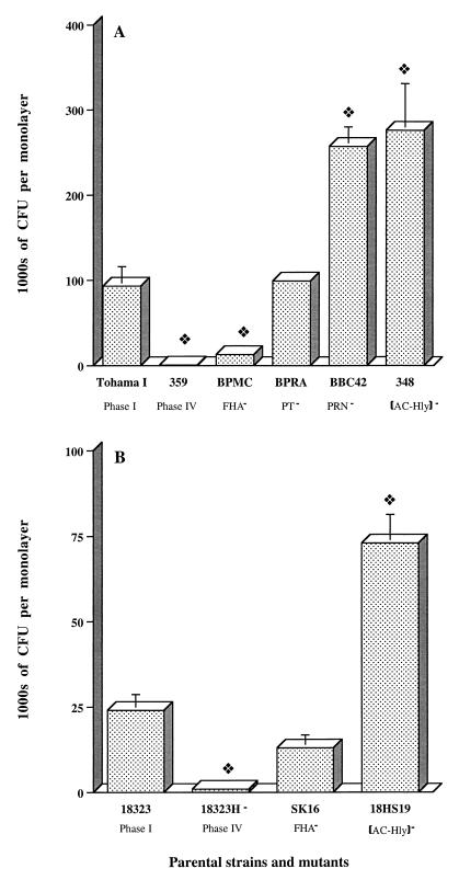
FIG. 2.
Invasion of HTE cell monolayers by B. pertussis mutants. Each B. pertussis strain (7 × 10⁶ CFU) was added to an individual well of a 24-well tissue culture plate, each of which contained 7 × 10⁴ epithelial cells. The invasion of HTE cells by B. pertussis was assayed as described in Materials and Methods. The values shown are means ± SEM in thousands of CFU recovered from gentamicin-treated monolayers in three to six experiments. The diamond symbol indicates P < 0.05 versus the parental B. pertussis strain, Tohama I (A) or 18323 (B).

