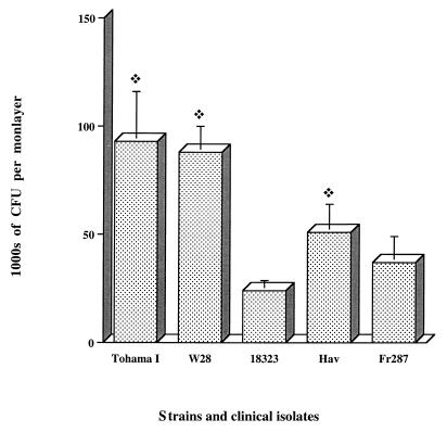
FIG. 4.
Invasion of HTE cell monolayers by B. pertussis isolates. Each B. pertussis strain or isolate (7 × 10⁶ CFU) was added to a separate well of a 24-well tissue culture plate each of which contained 7 × 10⁴ epithelial cells. The invasion of HTE cells by B. pertussis was assayed as described in Materials and Methods. The values shown are means ± SEM in thousands of CFU recovered from gentamicin-treated monolayers in three experiments. The diamond symbol indicates P < 0.05 compared to B. pertussis strain 18323.

