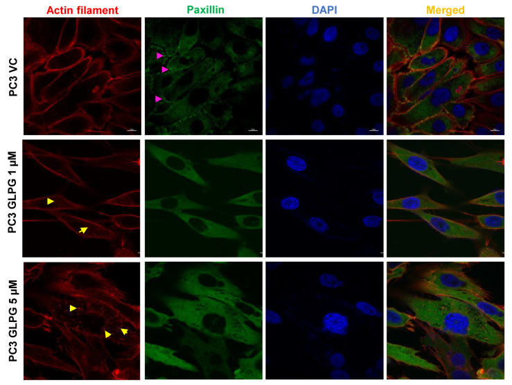
Figure 4.
MK5 regulates the formation of focal adhesions in the lamellipodial extensions of migratory PC3 cells. Immunostaining of paxillin along with rhodamine phalloidin–mediated actin staining to determine the localization of focal adhesions in PC3 cells treated with different doses of GLPG. Images are the representation of three independent experiments (n = 3). Scale bar = 10 µm. The pink arrow represents paxillin localization in the lamellipodial extensions in the vehicle control (VC) treated cells, while the yellow arrow represents the condensed actin foci in the basal surface of the GLPG-treated cells.

