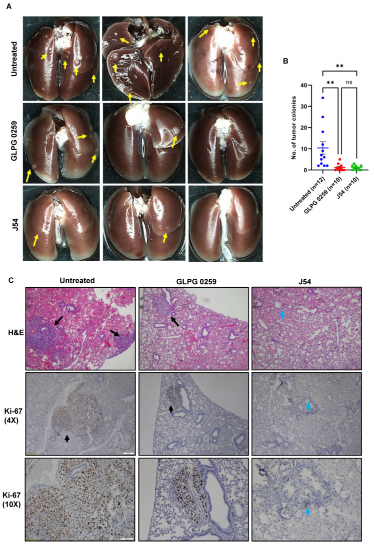
Figure 6.
TLK1 or MK5 inhibition impairs lung metastasis of PC3 cells in vivo. Five-week-old SCID beige mice were injected intravenously via the tail vein with 1 × 10⁶ parental PC3 cells and treated with either GLPG0259 or J54 intraperitoneally twice a week for 7 weeks. Lung metastases were compared among the untreated, GLPG-treated, or J54-treated mice. (A) Four representative images from each group are shown. (B) Data are presented as the number of metastatic colonies in the lung. (C) H&E and Ki-67 immunostaining shows lung nodules and proliferative tumor cells, respectively. Scale bar: 4× = 200 µm; 10× = 100 µm. One-way ANOVA, followed by Tukey’s post hoc analysis, was used for multiple group comparison: ** = p < 0.005; ns = not significant. Error bar represents standard error of the mean (SEM). The yellow and black arrows represent macroscopic nodules, whereas the blue arrows represent the presents of microscopic metastatic lesions.

