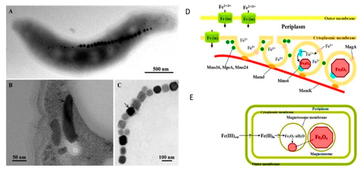
Figure 5.
Transmission electron microscope (TEM) images of magnetosomes and the magnetosome membrane. (A) TEM micrograph of a cell of Magnetospirillum magneticum strain AMB-1 deposited onto a Formvar-coated electron microscope grid showing a chain of cuboctahedral magnetosomes. (B) TEM micrograph of an ultrathin section of a cell of “Ca. Magnetoovum mohavensis” showing the magnetosome membrane (arrow) surrounding bullet-shaped magnetite crystals. (C) TEM micrograph of an extracted and purified magnetosome chain from a Magnetococcus marinus MC-1 cell showing prismatic magnetite crystals surrounded by the magnetosome membrane (arrow). (D) Scheme of the hypothesized mechanism of magnetite biomineralization. (E) Model for magnetite biomineralization in Magnetospirillum species [70,71]. Reprinted/adapted with permission from Refs. [70,71]. 2022, Elsevier B.V.

