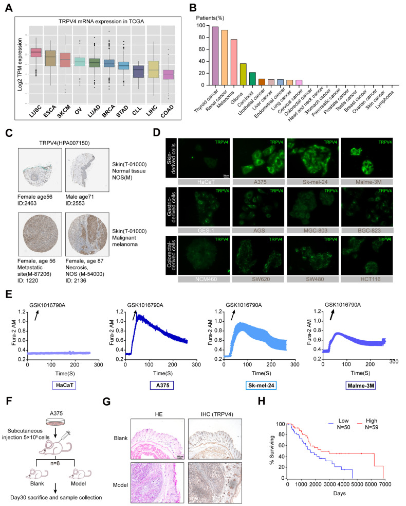
Figure 1.
Up-regulation of TRPV4 in melanoma. (A) TRPV4 mRNA expression level in different cancer tissues in TCGA. (B) TRPV4 protein expression level in different human cancerous and adjacent non-cancerous/normal tissue in terms of The Human Protein Atlas. (C) Strong immunoreactivity was observed in melanoma patients. (D) Representative images of TRPV4 expression (green) in multiple cells (Above: Skin-derived cell lines, HaCaT, A375, SK-mel-24, Malme-3M; Middle: Gastric-derived cell lines, GES-1, AGS, MGC-803, BGC-823; Below: Colorectal-derived cell lines, NCM460, SW620, SW480, HCT116). 400×. Scale bar, 20 μm. (E) Calcium imaging of HaCaT, A375, SK-mel-24, and Malme-3M cells treated with 2 nM GSK1016790A (n = 6, each group). (F) BALB/c-Nude mice were s.c. engrafted with 5 × 10⁶ A375 cells. (G) Representative images of HE and IHC (TRPV4) in tumor and normal skin tissues. 40×. Scale bar, 200 μm. (H) Survival cox regression data for TRPV4 from SKCM patients using OncoLnc (p-value = 0.0176). Data are expressed as means ± SD.

