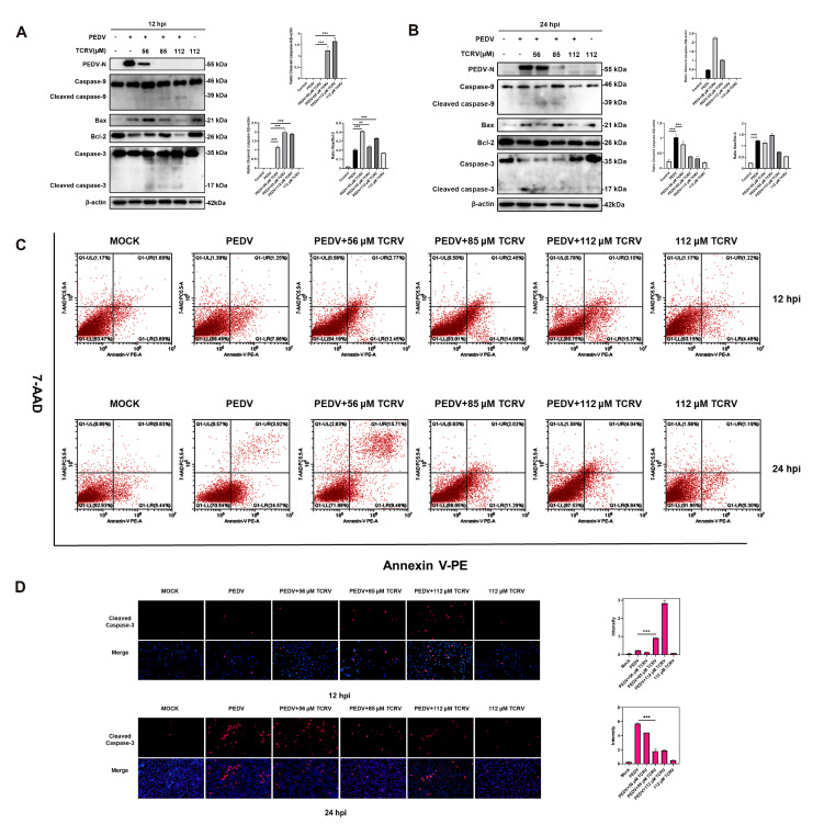
Figure 4.
The apoptosis induced by TCRV in PEDV-infected cells is different from PEDV-induced apoptosis. (A, B) Western blotting analysis of PEDV N, caspase-3, -9, Bax, and Bcl-2 proteins in PEDV-infected Vero cells treated with the indicated concentrations of TCRV at 12 hpi and 24 hpi. Results were presented as the ratio of band intensities of the target protein to β-actin. (C) PEDV-infected cells were treated with the indicated concentrations of TCRV at 12 hpi and 24 hpi. Apoptosis was analyzed using Annexin V-PE/7AAD staining, and the apoptotic cells were explored using flow cytometry. The Annexin V-positive cells were considered apoptotic cells. (D) The treatment of PEDV-infected cells with TCRV at concentrations of 56 µM, 85 µM, and 112 µM. Cells were processed for immunofluorescence using antibodies against cleaved caspase-3 protein and DAPI staining. Data from three independent experiments and error bars are presented as the mean ± SEM. ** p < 0.01, *** p < 0.001.

