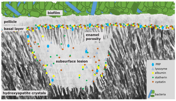
Figure 2.
Schematic illustration of a white spot lesion. It is characterized by a subsurface demineralization with an increased enamel porosity and porous outer tissues beneath. First, PRP and other components with inhibiting abilities protect the surface from further demineralization and also prevent crystal growth. Due to their size, they can only enter and diffuse into the deeper enamel layers at an intermediate level of caries lesion progression. At that state, the pores are wider, and small pellicle precursor and acid-resistant proteins might also be found inside the subsurface lesion. Consequently, differently sized proteins remain at different levels of the lesion, which they coat quickly during the demineralization process.

