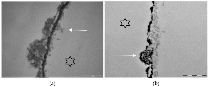
Figure 5.
Representative TEM-images visualize the pellicle ultrastructure. A subsurface pellicle can be detected after rinsing with fragaria vesca extract (a) and stannous fluoride (b) after 8 h of oral exposition [119,147]. Demineralized areas are filled with proteinaceous structures (arrows) and stannous ions (b). A higher electron density of the pellicle’s basal layer can be observed after rinsing with stannous ion containing agents. The enamel was removed during specimen processing and the former enamel side is marked by an asterisk.

