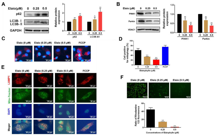
Figure 3.
Elaiophylin inhibits mitophagy and induces mitochondrial dysfunction in A549 cells. A549 cells were exposed to elaiophylin or FCCP (a mitophagy agonist) for 24 h. (A) The expression of autophagy-related proteins (LC3B and SQSTM1 (p62)) was assessed by Western blot analysis. (B) The expressions of mitophagy-related proteins (PINK1 and Parkin) were assessed by Western blot analysis. (C,D) A549 cells overexpressing Mito-Keima plasmid were treated with elaiophylin for 24 h. Mito-Keima (red fluorescence) was detected by a fluorescence microscope. FCCP, a mitophagy agonist, was used as a positive control. (E) Co-localization of mitochondria and lysosomes was assessed by MitoTracker (200 nM) and LAMP1 co-staining. (F) Mitochondrial membrane potential was assessed by rhodamine 123 staining. Data were expressed as means ± SD of three experiments and each experiment included triplicated repeats. * p < 0.05, ** p < 0.01 vs. control. Original Western Blots can be found at supplementary materials.

