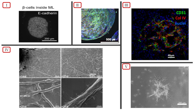
Figure 3.
(I) Spatial distribution of β cells inside EC spheroid, reported by Urbanczyk et al. [83] (licensed under CC BY 4.0). (II) Actin cytoskeletal organization and pre-vascular patterns in MSC/HUVEC spheroids encapsulated in collagen/fibrin hydrogels, reported by Heo et al. [79]. (III) Basal membrane highlighted using anti-collagen IV staining in vascularized spheroids, reported by Muller et al. [81] (licensed under CC BY 4.0). (IV) Scanning electron microscopy showcasing formation of endothelial cell tubes (indicated by arrows), reported by Chaddad et al. [85]. (V) Sprouting branches of co-cultured spheroids, reported by Kim et al. [84].

