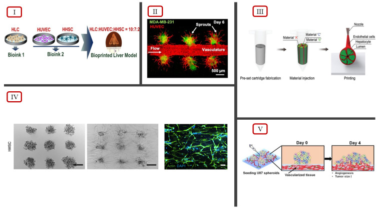
Figure 4.
(I) Bioink ratio to bioprint hepatic liver lobule model, illustrated by Janani et al. [88]. (II) Tumor angiogenesis observed using fluorescent imaging, reported by Dey et al. [90]. (III) Preset extrusion bioprinting technique to model hepatic lobule structure, illustrated by Kang et al. [89]. (IV) Self-organization of hMSCs via extrusion bioprinting using the BATE concept, reported by Brassard et al., scale bar 500 μm [87]. (V) Illustration of angiogenesis of MCTSs seeded on bioprinted vascularized tissue, reported by Han et al. [86] (licensed under CC BY 4.0).

