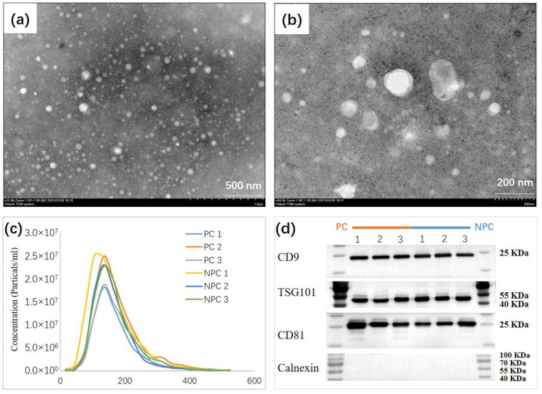
Figure 2.
Biochemical and Morphological Characterization of EVs. (a,b), TEM image of urine EV from selected individuals showing the typical exosome-round size (~150 nm) and morphology. (c) NTA of urine EV revealing a similar EV mode (~150 nm) between prostate cancer cases (n = 3), and non-prostate cancer controls (n = 3). (d) Representative immunoblots indicating that the EV isolated from the urine of selected individuals present enrichment of the EV markers CD9, TSG101, and CD81 but do not contain Calnexin used as control.

