Figure 2.
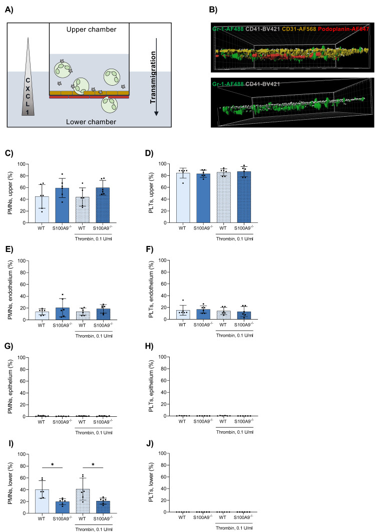
An in vitro murine bilayer model of primary alveolar endothelial and epithelial cells mimics the alveolar environment. Primary alveolar epithelial and endothelial cells were cultured on different sides of a TranswellTM insert. (A) Platelets and bone marrow-derived neutrophils (PMNs) were posited in the upper chamber and allowed to transmigrate along a CXCL1 gradient for 3 h at 37 °C with mild agitation (neutrophils = green, platelets = grey, endothelial cells = yellow, epithelial cells = red). After transmigration, TranswellTM membranes were cut out and treated with antibodies against Gr-1, CD41, CD31 and Podoplanin to stain neutrophils, platelets, endothelial and epithelial cells, respectively. Membranes were visualised via confocal microscopy (B). Neutrophil and platelet counts in the upper (C,D) and lower (I,J) TranswellTM compartment were determined by a Sysmex haemocytometer. Neutrophils and platelets attached to the endothelium (E,F) and epithelium (G,H) were counted in microscopic images. Data are mean ± SD, n = 6, * p < 0.05, two-way ANOVA.

